
Visual EM

Case Report of Untreated Pediatric Femoral Neck Fracture with Osteopenia
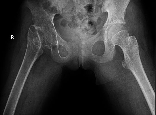
DOI: https://doi.org/10.21980/J8S92KOn her right hip radiograph, the patient was found to have a right femoral neck fracture with superior displacement of the intertrochanteric portion of the right femur. Moreover, the radiograph demonstrated diffuse osteopenia of the right hip and femur from chronic disuse as characterized by the increased radiolucency of the cortical bones compared to the left side.
High-Pressure Hand Injection Injury Case Report
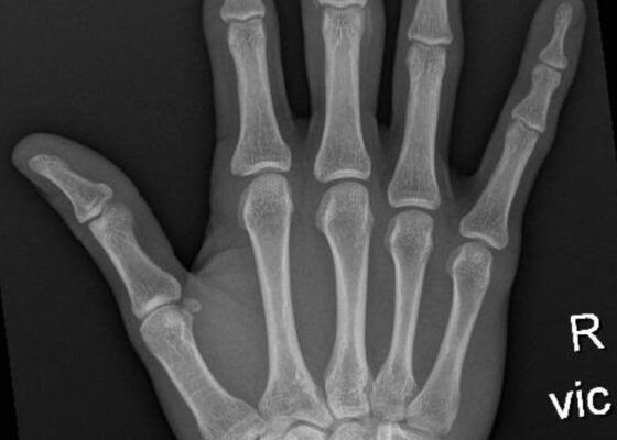
DOI: https://doi.org/10.21980/J8NM0PX-rays of his right hand revealed extensive infiltrates of the right distal and middle phalange without fractures or dislocations.

Loose PEG Tube Leading to Peristomal Leakage and Peritonitis
DOI: https://doi.org/10.21980/J8HS7TFrontal chest X-ray showed a large radiolucent area (pink highlighted area) underneath the diaphragm (yellow line) and on top of the liver (blue highlighted area) and spleen (green highlighted area) suggestive of pneumoperitoneum possibly caused by gastrointestinal perforation. This large radiolucent area can also be seen underneath the diaphragm in the lateral view chest X-ray. Computed tomography (CT) was not performed due to his physical exam findings and the significant positive findings on chest X-ray. Surgery was consulted and patient was taken emergently to the operating room.
Rapid Airway Narrowing Associated with Hodgkin’s Lymphoma
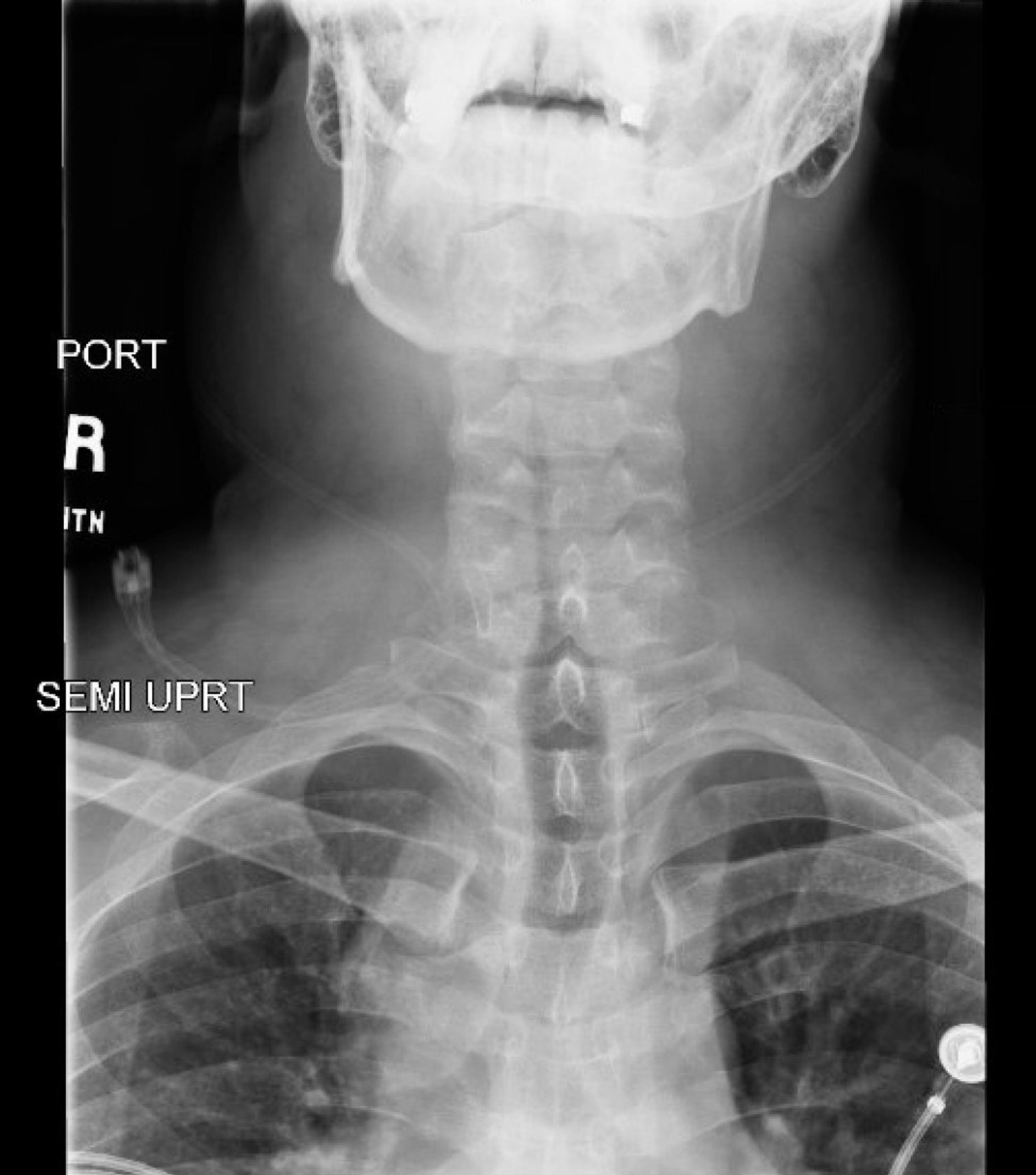
DOI: https://doi.org/10.21980/J86D3QNeck X-ray showed nonspecific significant prevertebral soft tissue swelling at the level of the cervical spine, with associated apparent thickening of the epiglottis (yellow arrow), diffuse soft tissue swelling of the neck (red arrows) and tracheal airway narrowing (light blue arrow). The computed tomography imaging of the neck was significant for multiple conglomerating pathological lymph nodes with a significant mass effect (orange arrows) compressing the right internal jugular vein (green arrow).

Fitz Hugh Curtis Case Report
DOI: https://doi.org/10.21980/J82K9GA sagittal view from computed tomography (CT) of the abdomen and pelvis demonstrated fat stranding beneath the inferior margin of the liver (outlined in red). The axial view showed fat stranding adjacent to the ascending colon without significant colon wall thickening (arrow). Fat stranding can occur as a hazy increased attenuation (brightness) or a more distinct reticular pattern.

Ascending Thoracic Aortic Dissection: A Case Report of Rapid Detection Via Emergency Echocardiography with Suprasternal Notch Views
DOI: https://doi.org/10.21980/J8WW6WVideo of parasternal long-axis bedside transthoracic echocardiogram: The initial images showed grossly normal left ventricular function, and no pericardial effusion or evidence of cardiac tamponade. However, the proximal aorta beyond the aortic valve was poorly-visualized in this window.

Hemorrhagic Renal Cyst
DOI: https://doi.org/10.21980/J8C92VBedside renal ultrasound demonstrated a right renal cyst with echogenic debris consistent with a hemorrhagic cyst (red arrow). In addition, a computed tomography (CT) scan of the abdomen and pelvis revealed a 4mm non-obstructing right renal stone and bilateral renal cysts. The CT also confirmed the ultrasound finding of a right renal cyst with mild perinephric stranding possibly consistent with a hemorrhagic cyst.
Meckel’s Diverticulum Causing Small Bowel Intussusception in Third Trimester Pregnancy
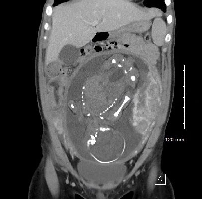
DOI: https://doi.org/10.21980/J87H19A CT scan was obtained which demonstrated distal small bowel intussusception with focal dilation suggestive of a small bowel obstruction in a pregnant female in her third trimester of pregnancy. The fetus can be seen in the uterus. The yellow arrow identifies the area of small bowel intussusception shown by telescoping intestines with associated bowel wall edema.