
JETem
  • Home
  • About
    • Aim and Scope
    • Our Team
    • Editorial Board
    • FAQ
  • Issues
    • Current Issue
    • Ahead of Print
    • Past Issues
  • Visual EM
    • Latest Visual EM
    • Search Visual EM
    • Thumbnail Library
  • For Authors
    • Instructions for Authors
    • Submit to JETem
    • Photo Consent
    • Policies
      • Peer Review Policy
      • Copyright Policy
      • Editorial Policy, Ethics and Responsibilities
      • Conflicts of Interest & Informed Consent
      • Open Access Policy
      • Acceptance Fee
  • For Reviewers
    • Instructions for JETem Reviewers
    • Interested in Being a JETem Reviewer?
  • Topic
    • Abdominal/Gastroenterology
    • Administration
    • Board Review
    • Cardiology/Vascular
    • Certifying Exam Cases
      • Clinical Decision-Making
      • Prioritization
      • Communication
      • Procedure & Ultrasound
    • Clinical Informatics, Telehealth and Technology
    • Dermatology
    • EMS
    • Endocrine
    • ENT
    • Faculty Development
    • Genitourinary
    • Geriatrics
    • Hematology/Oncology
    • Infectious Disease
    • Miscellaneous (stats, etc)
    • Neurology
    • Ob/Gyn
    • Ophthalmology
    • Orthopedics
    • Pediatrics
    • Pharmacology
    • Procedures
    • Psychiatry
    • Renal/Electrolytes/Nutrition
    • Respiratory
    • Social Determinants of Health
    • Toxicology
    • Trauma
    • Ultrasound
    • Urology
    • Wellness
    • Wilderness
  • Modality
    • Certifying Exam Practice Cases
      • Clinical Care Cases
      • Communication & Procedural Cases
      • Old School Oral Board Cases
    • Curricula
    • Innovations
    • Lectures
    • Podcasts
    • Simulation
    • Small Group Learning
    • Team Based Learning (TBL)
    • Visual EM
  • Contact Us

Posts by JETem

Posterior Reversible Encephalopathy Syndrome and Eclampsia

Kristina Jacomino, MD*, Kevin Tomecsek, MD*, Andrew Little, DO* and Mary Mclean, MD*

DOI: https://doi.org/10.21980/J8H64T Issue 10:3[mrp_rating_result]
At the end of this oral boards session, examinees will be able to: 1) demonstrate familiarity with the structured interview oral board format and case play; 2) recognize the history and exam features concerning for PRES and eclampsia; 3) order appropriate diagnostic workup for postpartum and hypertensive emergencies including eclampsia and PRES; 4) understand treatment options for the management of eclampsia (intravenous [IV] magnesium sulfate, IV antihypertensive therapy, and emergent consultation with an obstetrician [OB/GYN]); 5) understand threshold for taking control of airway in patients with eclampsia; 6) understand indications for ordering brain imaging in patients with eclampsia and altered mental status; and 7) demonstrate effective communication with treatment team/family members as well as correct disposition of the patient to a higher level of care (intensive care unit [ICU]).
Certifying Exam CasesNeurologyOb/Gyn
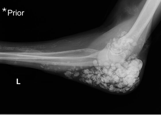
Calcinosis Cutis. Prior Elbow XR Lateral. JETem 2025

Metastatic Calcinosis Cutis in the Emergency Department: A Case Report

Christian Hernandez-Zegada, MD*, Holly Conger, MD* and Brian Milman, MD*

DOI: https://doi.org/10.21980/J87Q00 Issue 10:3[mrp_rating_result]
X-ray imaging was obtained of the left elbow and showed soft tissue calcium deposits. Radiology stated, “massive periarticular calcinosis of renal failure obscures fine osseous detail. Several of the largest calcifications have decompressed since the prior exam and may contribute to the drainage observed clinically. Superimposed infection is not excluded.” X-rays with an asterisk are the comparison images from two months previous to the visit. Areas of decompression are highlighted in blue demonstrating that some of the larger calcified nodules are no longer present.
Visual EMDermatologyRenal/Electrolytes
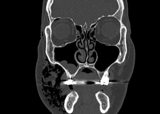
Dental Free Air CT Coronal Unannotated. JETem 2025

A Case Report of Facial Swelling and Crepitus Following a Dental Procedure

Shady Mikhail, MD*, George Mina, MD^, Alisa Wray MD, MAEd† and Danielle Matonis, MD†

DOI: https://doi.org/10.21980/J83W8H Issue 10:3[mrp_rating_result]
Given the physical exam findings of crepitus on the right neck up to the right lower eyelid, a maxillofacial CT scan without contrast was performed. It revealed diffuse subcutaneous air within the soft tissues of the face and neck and free air within the pre-septal soft tissue of the right eye, appearing as hyperlucent (dark) areas on CT within the soft tissue planes (blue outline). It showed no evidence of post-septal free air. A single-view chest X-ray was also performed and was unremarkable except for incompletely imaged soft tissue gas in the right lower neck (blue outline). On flexible fiberoptic laryngoscopy performed by ENT, the oropharynx appeared diffusely edematous and narrowed.
Visual EMENT
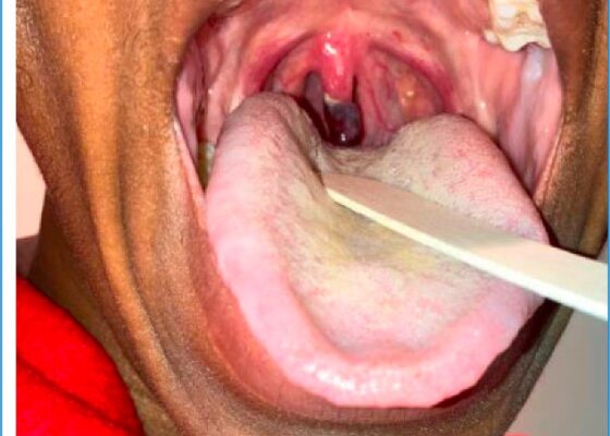
Uvular Necrosis Photo. Unannotated. JETem 2025

Case Report of Post-Operative Uvular Necrosis Following Intubation

Laryssa Patti, MD*, Jal Trivedi, MD^ and Mary Rometti, MD†

DOI: https://doi.org/10.21980/J8065J Issue 10:3[mrp_rating_result]
The distal portion of her uvula was necrotic with a clear demarcation approximately halfway up the uvula. She had no trauma to the anterior oropharyngeal structures, tonsils, or adenoids. There were no lesions to the hard or soft palate. She had no carotid bruits or thrills, and no tenderness over the anterior portion of the neck.
Visual EMENT
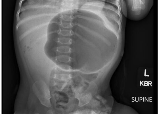
Incarcerated Gastric Volvulus and Splenic Herniation. Abdominal Radiograph. Unannotated. JETem 2025

Case Report of Incarcerated Gastric Volvulus and Splenic Herniation in Undiagnosed Congenital Diaphragmatic Hernia in an Infant

Kate R Gelman*, Torren A Kalaskey*and Federico G. Seifarth, MD ^

DOI: https://doi.org/10.21980/J8VD27 Issue 10:3[mrp_rating_result]
An upper gastrointestinal series (UGI) showed an enteric tube with its tip in the stomach and side-port in the esophagus. There was a large amount of air in the stomach and a small volume of scattered distal bowel gas. The tip of an enteric tube was seen in the stomach (red arrow). Contrast partially filled the stomach, and the greater curvature was visualized superior to the lesser curvature in the left upper quadrant (blue arrow). The body of the stomach was herniated into the right chest through a Bochdalek hernia (blue star). There was a large amount of air in the stomach and a small volume of scattered distal bowel gas. These findings were consistent with mesenteroaxial gastric volvulus.
Visual EMAbdominal/GastroenterologyPediatrics
Mycoplasma Associated Acute Transverse Myelitis. MRI. Unannotated. JETem 2025

Beware of the Pediatric Limp: A Case of Mycoplasma Associated Acute Transverse Myelitis

Michael Neff, MD*, Nicholas Xie, MD^, Joseph Fong, MD† and Gregory Podolej, MD**

DOI: https://doi.org/10.21980/J8QQ1Q Issue 10:3[mrp_rating_result]
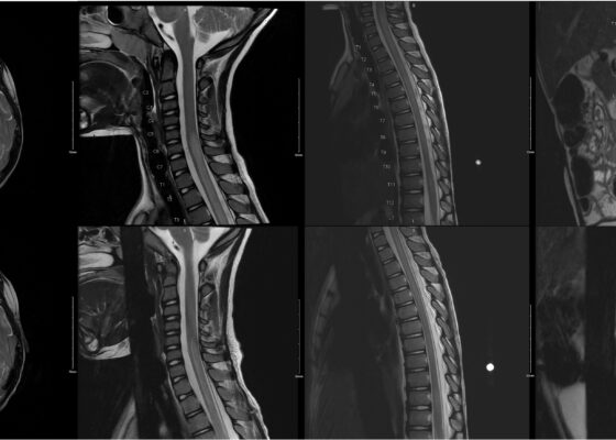
An MRI with contrast, T2 sequence was performed. In Figures a-d, the MRI of the patient’s brain and spinal cord on admission shows abnormal signals in the patient’s pons (lack of symmetrical gray-white differentiation on cross-section) along with hyperintensity (sagittally shown as brightness in what should be homogenously intense spinal cord) and significant central cord edema (with swelling seen as increased width) starting from C5 and continuing to the conus medullaris around L1/L2.
Visual EMNeurologyPediatrics
Calciphylaxis Photo LLE. JETem 2025 1

A Case Report of Calciphylaxis

Kim Hoang, BS*, Tien Lu, MD^, Alex Dang, MD^, Danielle Matonis, MD^

DOI: https://doi.org/10.21980/J8KW8V Issue 10:3[mrp_rating_result]
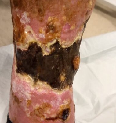
On arrival for this visit, the patient was nontoxic appearing with stable vital signs. The physical exam was notable for deep, ulcerated, bilateral anterior leg wounds with purulent drainage and large areas of eschar (see photographs).
Visual EMDermatologyInfectious DiseaseRenal/Electrolytes
Iatrogenic Bowel Perforation Following Dental Procedure. CT. Unannotated. JETem 2025

Case Report: Iatrogenic Bowel Perforation Following Dental Procedure

Claire DeLong, DO* and Frederick Fiesseler, DO*

DOI: https://doi.org/10.21980/J8CD38 Issue 10:3[mrp_rating_result]
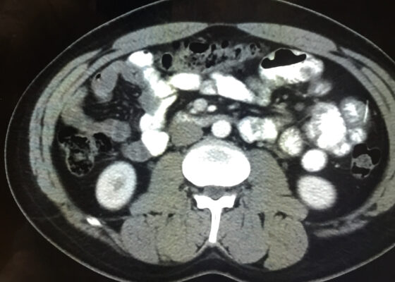
The patient’s abdominal CT demonstrated a metallic foreign body in the left side of the abdomen within the small bowel, without surrounding induration or abscess. Radiology questioned whether the metallic foreign object perforated the bowel. Seen in the cross-sectional CT image, there is a hyperdense linear structure transversing the small intestinal wall, given that a portion of the structure was located outside of the lumen of the bowel.
Visual EMAbdominal/Gastroenterology
«‹12›»
Page 12 of 91

JETem is an online, open access, peer-reviewed, journal-repository for EM educators. We are PMC Indexed.

Most Viewed

  • Simple Auricular Hematoma (“Cauliflower Ear”) Task Trainer
  • The Three Breath Method: Teaching Coping Mechanisms for Processing Patient Death
  • Clinical Decision Making: Acute Ischemic Stroke with Large Vessel Occlusion
  • PRIMER: Premedical Introduction to Mentored Emergency Research: Connecting Undergraduate Assistants and Emergency Medicine Residents in a Community Hospital Setting
  • Dangerous Dysrhythmias in the ED: An Interactive EKG Workshop for Emergency Medicine Clerkship Learners

Visit Our Collaborators


Creative Commons Licence
This work is licensed under a Creative Commons Attribution 4.0 International License.

About

Education

Learners should benefit from active learning. JETem accepts submissions of team-based learning, small group learning, simulation, podcasts, lectures, innovations, curricula, question sets, and visualEM.

Scholarship

We believe educators should advance through the scholarship of their educational work. JETem gives educators the opportunity to publish scholarly academic work so that it may be widely distributed, thereby increasing the significance of their results.

Links

  • Home
  • Aim and Scope
  • Current Issue
  • For Reviewers
  • Instructions for Authors
  • Contact Us

Newsletter

Sign up to receive updates from JETem regarding newly published issues and findings.

Copyright Creative Commons Attribution 4.0 International