Latest Articles
A Low Cost Escharotomy Simulation Model for Residency Education
DOI: https://doi.org/10.21980/J8D34VBy the end of this educational session, the learner will be able to: 1) Understand the indications for an escharotomy. 2) List equipment needed to perform an escharotomy. 3) Demonstrate how to perform an escharotomy. 4) Perform an escharotomy and experience the sensation of cutting through simulated burned tissue. 5) Understand post-escharotomy management and referral to specialist.
Acute Hemolytic Transfusion Reaction
DOI: https://doi.org/10.21980/J88D2ZBy the end of this simulation session, the learner will be able to: 1) Recognize the clinical signs and symptoms associated with transfusion reactions. 2) Discuss necessary systems-based management of potential transfusion reactions, such as notifying the blood bank and evaluating to see if another patienta accidentally received a wrong unit of blood. 3) Discuss the management of various transfusion reactions. 4) Appropriately disposition the patient to an intensive care unit (ICU) or stepdown unit. 5) Effectively communicate with team members and nursing staff during the resuscitation of a critically ill patient.
In-Flight Emergency: Altered Mental Status Secondary to Hypoglycemia
DOI: https://doi.org/10.21980/J84M01By the end of this simulation session, learners will be able to: 1) Discuss the challenges associated with in-flight emergencies. 2) List what is available in United States (US) commercial airline medical kits. 3) Discuss an appropriate differential diagnosis for a patient with altered mental status (AMS). 4) Demonstrate appropriate treatment of hypoglycemia. 5) Describe possible complications in diabetic patients with insulin pumps when flying. 6) Review the legal ramifications of responding to an in-flight emergency.
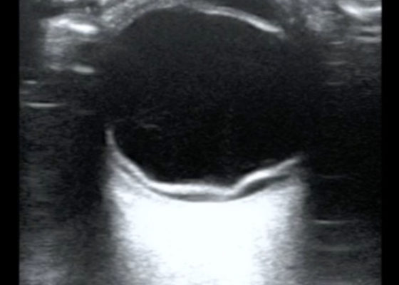
Bedside Ultrasound of Retinal Detachment in a 19-year-old
DOI: https://doi.org/10.21980/J80W6TThe ocular point of care ultrasound (POCUS) utilizing a high frequency linear probe shows a retinal detachment (RD) with a thick, hyperechoic undulating membrane in the vitreous humor that is anchored at the ora serrata anteriorly and the optic disc posteriorly. Note that the retina is detached all the way to the optic disc making it "mac off." The macula, and more specifically the fovea, is located in the central retina and contains a high concentration of cone photoreceptors responsible for central, high resolution, color vision. In a "mac on" RD, the retina detaches in the periphery but remains intact centrally. This is an ophthalmologic emergency and timely diagnosis and intervention can be vision saving. This patient also has evidence of a posterior vitreous hemorrhage which has a characteristic swirling appearance with kinetic exam on real-time imaging. The detached vitreous body is not as well defined and is not anchored posteriorly to the optic disc.
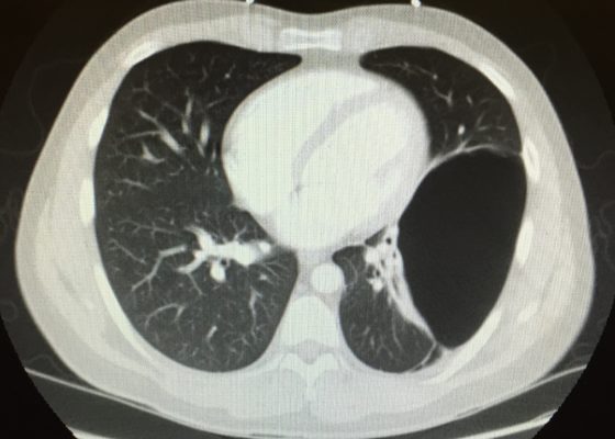
Bullous Emphysema
DOI: https://doi.org/10.21980/J8W62GThe upright chest X-ray shows a large lucent area in the left lower lung field without lung markings, with associated curvilinear opacities (yellow arrows) consistent with a large air-filled bulla. The bulla is large enough to compress adjacent lung tissue as shown by the visible pleural line (blue line). The discontinuity of the pleural line and presence of lung markings superiorly makes these findings more consistent with bulla than pneumothorax. The chest computed tomography (CT) confirmed a large left hemithorax bulla.
The Role of Chest X-Ray and Bedside Ultrasound in Diagnosing Pulmonary Bleb versus Pneumothorax
DOI: https://doi.org/10.21980/J8MP7QThe patient was evaluated with bedside ultrasound for concern of possible pneumothorax. Imaging of the left lung with M-mode demonstrated a “sea shore” sign showing a wavy pattern below the pleural line caused by lung sliding as well as “comet tail” artifact caused by from the deep pleura. However, there was no lung sliding on the right shown by a lack of “comet tail” artifact and a “bar code” sign where M-mode shows straight lines throughout the image, this is caused by lack of motion below the pleura. This lack of lung sliding is consistent with possible pneumothorax or bleb.
A two-view chest X-ray (CXR) revealed absent lung parenchyma in the right lung similar to a large pneumothorax (see red outline). Electronic medical record chart review revealed previous CXRs with similar findings. This patient was determined to have an acute COPD exacerbation with chronic blebs, but no pneumothorax.


